Single-Dose CaBP4 Gene Therapy Rewired Adult Dog Retinas and Rebuilt the Synaptic Layer That Never Formed

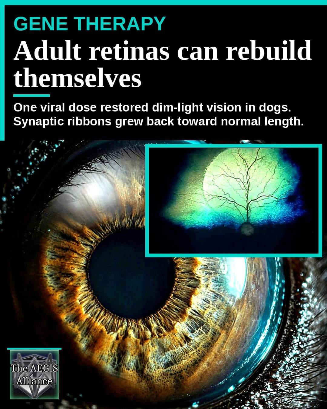
A dog born with a broken copy of CaBP4 does not simply lose a chemical. The first synapse of the retina never finishes building. On September 28, 2026, Michigan State University ha descritto una terapia genica che ha fatto più che cambiare quel segnale negli animali adulti. patch trattate di retina addensato uno strato che era rimasto sottile dopo lo sviluppo, e nastri sinaptici all'interno delle cellule di luce-sensamento è cresciuto fino a quando non erano difficili da dire da quelli in occhi sani.
Questo è un reclamo diverso da Luxturna, il farmaco del 2017 che fornisce un enzima mancante alle cellule retiniche che sono ancora cablate insieme. Qui il cablaggio stesso era stato lasciato incompiuto. Il documento, nel numero del 10 settembre 2026 Molecular Therapy Advances, calls the result plasticity in an adult mammalian synapse.
A Typo in the Retinal Blueprint Stopped the First Synapse
CaBP4 codifica la proteina che lega il calcio 4. Al terminale del fotorecettore, il calcio consente a un nastro di rilascio di sinapsi glutammato sulle cellule bipolari. Quando la proteina fallisce, quella transazione fallisce con esso. Nelle persone il gene è di solito scritto CABP4, e le mutazioni in entrambe le copie sono una causa di cecità notturna stazionaria congenita incompleta, con scarsa acuità, problemi in luce debole, e un elettroretinogramma in cui i fotorecettori rispondono mentre la cella successiva rimane tranquilla. A 2025 Journal of Translational Medicine review shows how often gene-therapy plans for this family of disorders stall on which cell to treat.
Billie Beckwith-Cohen, un oftalmologo veterinario nello Stato del Michigan, ha confrontato la mutazione ad un typo in un piano di costruzione. "Si può essenzialmente discutere le mutazioni del gene retinico come un typo in un'impronta che rende le istruzioni incomprensibili al sistema, con conseguente progettazione difettosa e successiva perdita di visione", ha detto. "La nostra terapia fornisce essenzialmente nuove istruzioni per il segmento misspelled, come un editor."
I ricercatori hanno trovato una mutazione spontanea di CaBP4 in fruste la cui sire e diga ha avuto origine in Brasile, poi ha mantenuto una colonia riproduttiva in East Lansing. Un occhio di cane, grande e ricco di coni, è uno stand-in più vicino per un paziente che una retina del topo, motivo per cui Simon Petersen-Jones ha usato la canina ereditata malattia retinica come ponte alle prove umane, anche prima Lavoro CNGB1.

What the CaBP4 Mutation Did to the Outer Plexiform Layer
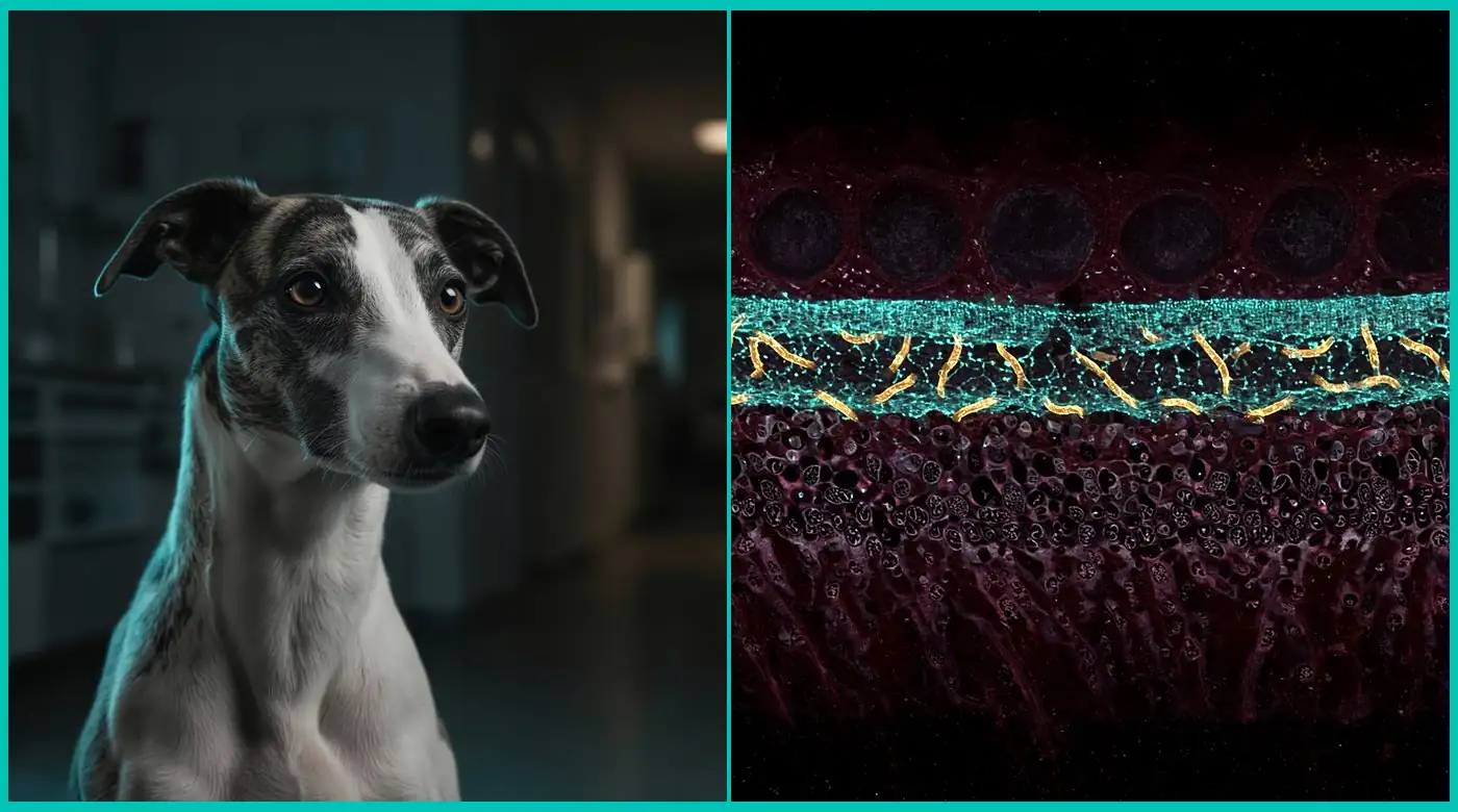
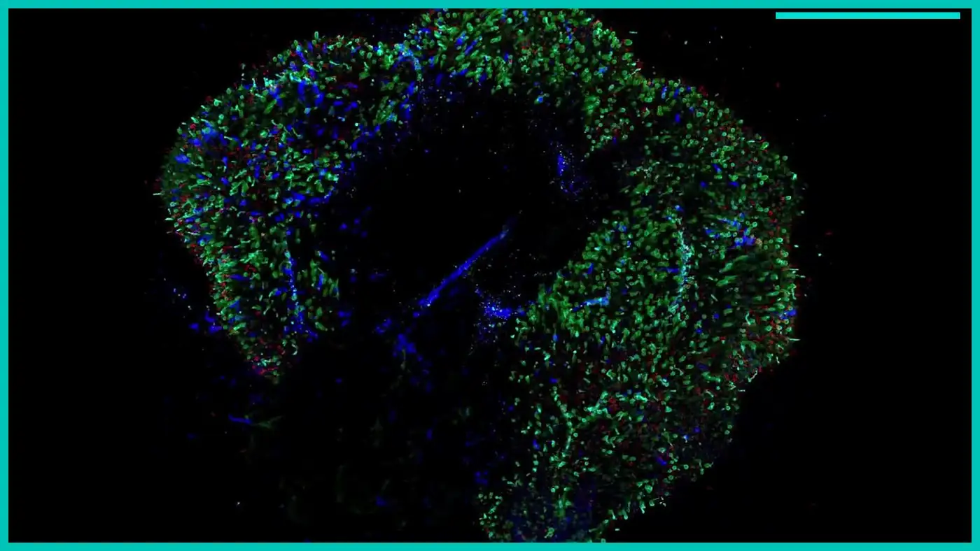
Lo strato plexiforme esterno, o OPL, è la sottile banda sinattica dove vivono quelle connessioni. Nei cani mutanti si formava a malapena. La carta riporta un OPL di 2,87 ± 0,20 micrometri negli animali carenti di CaBP4, contro 8,27 ± 0,15 micrometri nei controlli non colpiti. I nastri sinaptici sono stati immatura a 227 ± 14 nanometri, rispetto a 642 ± 27 nanometri in retina di tipo selvaggio. La b-wave, la firma elettrica dell'attività bipolare-cellula, era assente, e la visione era peggiore al buio.
Nel corso del tempo la retina esterna si è assottigliata, quindi le cellule un trattamento successivo avrebbe bisogno di iniziare a scomparire. Gli autori chiamano la colonia il primo modello naturale di grande animale mutante CaBP4 che ricapitola parti della malattia umana.
One AAV Dose Rebuilt Ribbons That Had Stayed Immature
Il trattamento è stato l'aumento del gene, non la modifica del gene. Un chirurgo ha posto un virus adeno-associated, AAV8 con una variante 733 capsid, sotto la retina. Un promotore GRK1 ha guidato sia la sequenza di codifica canina che quella umana. Dose corse da 2 × 10¹⁰ a 8,5 × 10¹¹ vector genomes per eye. Both versions rescued the animals, with no meaningful difference between the dog gene and the human gene.
La sorpresa è stata la ricostruzione. OPL trattato misura 6,8 ± 0,5 micrometri, contro 1,7 ± 0,8 micrometri nelle regioni non trattate della retina colpita. Gli occhi sani si sedevano vicino a 8,2 micrometri, quindi lo strato riparato era ancora leggermente sottile, ma la maggior parte dello spessore mancante era tornato. I nastri nei tessuti trattati hanno raggiunto 604 ± 15 nanometri, contro circa 245 ± 10 nanometri lasciati non trattati. La lunghezza trattata non era statisticamente diversa dai nastri di tipo selvaggio.
La carta descrive anche nuove triadi sintattiche, i contatti tra un fotorecettore, una cellula bipolare e una cellula orizzontale, e il ritorno di proteine come GPR179 a destra indirizzo. Beckwith-Cohen ha detto che il lavoro ha mostrato tre cambiamenti strutturali indipendenti che sostengono la plasticità nella retina adulta. "Non solo sono stati aggiunti nuovi componenti, ma le anomalie preesistenti sono state riparate."
Michigan State ha chiamato il progetto il prodotto di circa dieci anni nel laboratorio Petersen-Jones, dal trovare la mutazione alla costruzione del vettore. I coautori includono scienziati della Pontifícia Universidade Católica do Paraná e dell'Universidade Federal do Paraná, che è adatto. La storia genetica dei cani è iniziata in Brasile.
Why a Spontaneous Whippet Disease Beats a Mouse Knockout
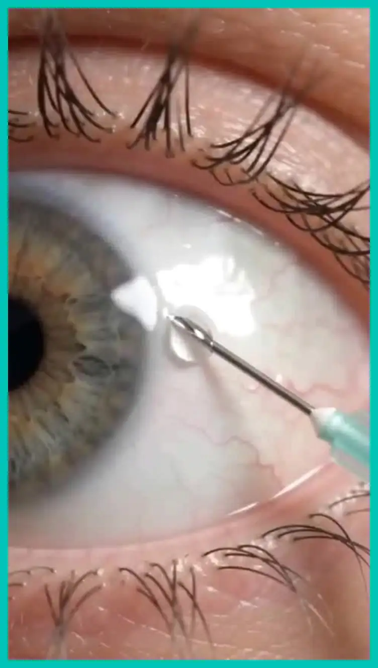
Un'iniezione subretinale in un occhio di cane è una prova per l'operazione che un bambino avrebbe affrontato, e una malattia ereditaria è un test più equo di un topo knockout. Il documento dice che il ripristino della regolazione del calcio era necessario per il rimodellamento.
Retinal gene therapy already has a famous dog in its origin story. Lancelot, treated at the University of Pennsylvania, helped carry RPE65 research to Luxturna, approved in 2017. Luxturna, or voretigene neparvovec, gives surviving cells a working RPE65 gene so the visual cycle can run again. Boston Children’s Hospital limits it to people with mutations in both copies of that gene and enough retina left to respond. It does not ask an adult eye to build a synaptic layer development skipped.
A sickle cell gene therapy such as Lyfgenia changes how blood cells make hemoglobin. It does not regrow a neural circuit. If an adult outer plexiform layer can expand once calcium signaling returns, the old assumption that mammalian retinal synapses are finished at birth looks weaker.

The Repair Is Real and It Is Still Regional
The virus only reaches the retinal bleb the surgeon creates, and a full-field electroretinogram still averages in untreated retina, so recovered waveforms will not match a healthy eye. The paper reports a returned scotopic b-wave of 91 ± 18 at a dim flash of 0.01 candela-seconds per square meter. Bright-light gains were not always significant in young dogs that still had some day vision. If photoreceptors had already died, little remained to rescue. This retina could remodel a synapse. It could not replace a cell that was gone.
Structural recovery held for more than two years in animals tracked that far. Dogs treated at 28 months of age, already adult, were followed for 18 months. One animal treated at four and six months was followed to 44 months. That is a durability finding in dogs, not a promise for people.
The outer plexiform layer could be expanded beyond the thickness seen in the young mutant dogs before therapy, the authors write, “illustrating that photoreceptor pedicles and bipolar cell dendrites can extend or retract to establish a normal retinal layer, expanding a synaptic space that had not previously developed.”
What This Suggests Beyond a Rare Night-Blindness Gene
CABP4 disease is rare in people and in dogs. This is not a treatment for macular degeneration, glaucoma, or ordinary sight loss from age. In this model, restoring calcium control at the first synapse let an adult circuit finish a job development had dropped. The abstract calls that restoration requisite for retinal plasticity and says the model paves the way for a cure of CaBP4 blindness. The discussion is stricter. It offers a foundation for studying the human condition, not a trial protocol.
CaBP4 sits on the photoreceptor side of the synapse, which is why a GRK1-driven virus, built to enter rods and cones, was a logical vehicle. Disorders that live in the bipolar cell will need a different address. A transplanted heart tends to take on the recipient’s biological age rather than rewind it. These retinas did not become embryonic either. They finished a missing piece of their own architecture once the missing protein arrived. Dose, immune response, surgical coverage, and whether children with CABP4 mutations still have living photoreceptors will decide if a trial is ethical. The human coding sequence already works in the dog. For readers following health reporting e il scienza scrivania, that sequence is the bridge, and the adult outer plexiform layer’s willingness to be built is the surprise.
The study is Gene therapy induces synaptic ribbon maturation, synaptogenesis and vision recovery in an adult dog model of retinal degeneration, article 201782 in Molecular Therapy Advances.