Single-Dose CaBP4 Gene Therapy Rewired Adult Dog Retinas and Rebuilt the Synaptic Layer That Never Formed
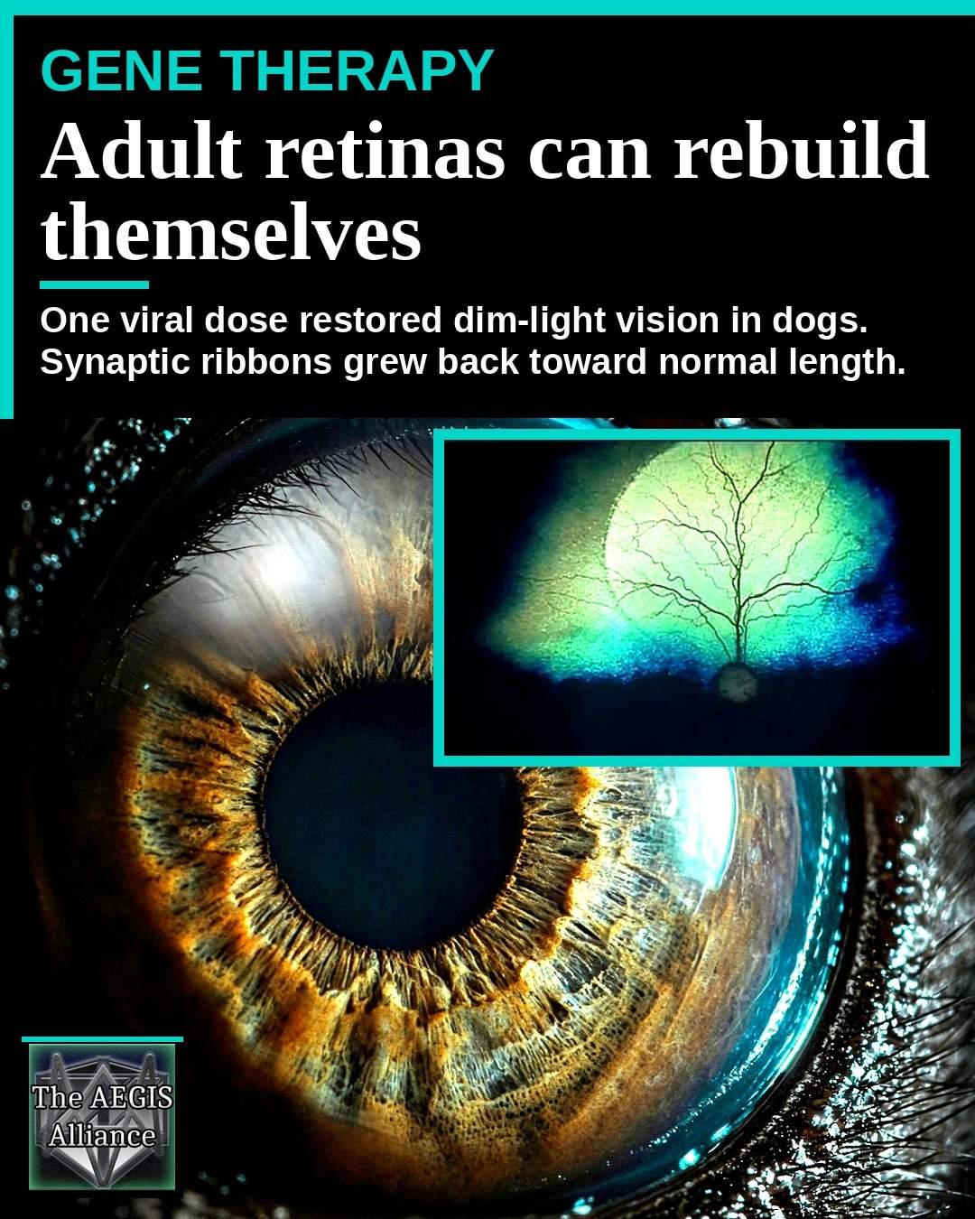
A dog born with a broken copy of CaBP4 does not simply lose a chemical. The first synapse of the retina never finishes building. On September 28, 2026, Michigan State University described a gene therapy that did more than switch that signal back on in adult animals. Treated patches of retina thickened a layer that had stayed thin since development, and synaptic ribbons inside light-sensing cells grew until they were hard to tell from those in healthy eyes.
That is a different claim from Luxturna, the 2017 drug that supplies a missing enzyme to retinal cells that are still wired together. Here the wiring itself had been left unfinished. The paper, in the September 10, 2026 issue of Molecular Therapy Advances, calls the result plasticity in an adult mammalian synapse.
A Typo in the Retinal Blueprint Stopped the First Synapse
CaBP4 encodes calcium-binding protein 4. At the photoreceptor terminal, calcium lets a ribbon synapse release glutamate onto bipolar cells. When the protein fails, that transaction fails with it. In people the gene is usually written CABP4, and mutations in both copies are one cause of incomplete congenital stationary night blindness, with poor acuity, trouble in dim light, and an electroretinogram in which photoreceptors respond while the next cell stays quiet. A 2025 Journal of Translational Medicine review shows how often gene-therapy plans for this family of disorders stall on which cell to treat.
Billie Beckwith-Cohen, a veterinary ophthalmologist at Michigan State, compared the mutation to a typo in a building plan. „One can essentially discuss the mutations in the retinal gene as a typo in a blueprint that makes the instructions incomprehensible to the system, resulting in a faulty design and subsequent vision loss,“ she said. „Our therapy essentially provides new instructions for the misspelled segment, like an editor.“
Researchers found a spontaneous CaBP4 mutation in whippets whose sire and dam originated in Brazil, then kept a breeding colony in East Lansing. A dog eye, large and rich in cones, is a closer stand-in for a patient than a mouse retina, which is why Simon Petersen-Jones has used canine inherited retinal disease as a bridge to human trials, including earlier CNGB1 work.
What the CaBP4 Mutation Did to the Outer Plexiform Layer
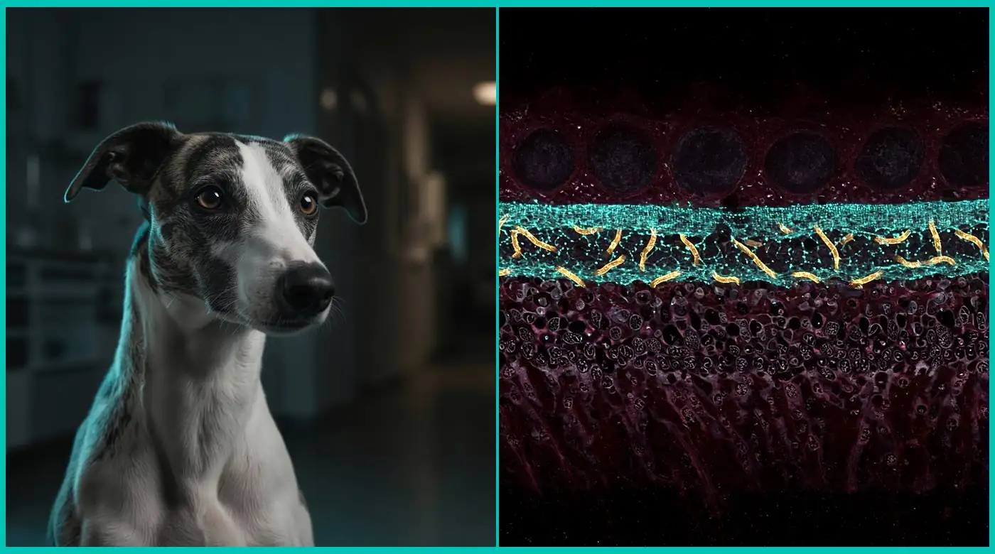
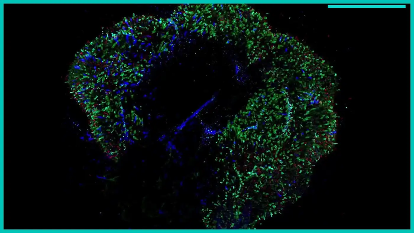
The outer plexiform layer, or OPL, is the thin synaptic band where those connections live. In the mutant dogs it barely formed. The paper reports an OPL of 2.87 ± 0.20 micrometers in CaBP4-deficient animals, against 8.27 ± 0.15 micrometers in unaffected controls. Synaptic ribbons were immature at 227 ± 14 nanometers, compared with 642 ± 27 nanometers in wild-type retina. The b-wave, the electrical signature of bipolar-cell activity, was absent, and vision was worse in the dark.
Over time the outer retina thinned, so cells a later treatment would need began to disappear. The authors call the colony the first naturally occurring large-animal model of mutant CaBP4 that recapitulates parts of the human disease.
One AAV Dose Rebuilt Ribbons That Had Stayed Immature
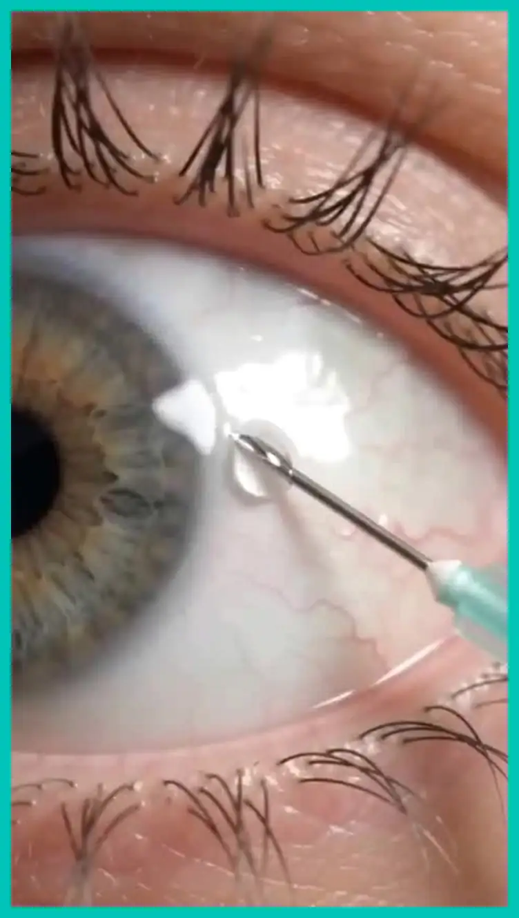
The treatment was gene augmentation, not gene editing. A surgeon placed an adeno-associated virus, AAV8 with a 733 capsid variant, under the retina. A GRK1 promoter drove either the canine coding sequence or the human one. Doses ran from 2 × 10¹⁰ to 8.5 × 10¹¹ vector genomes per eye. Both versions rescued the animals, with no meaningful difference between the dog gene and the human gene.
The surprise was reconstruction. Treated OPL measured 6.8 ± 0.5 micrometers, against 1.7 ± 0.8 micrometers in untreated regions of affected retina. Healthy eyes sat near 8.2 micrometers, so the repaired layer was still slightly thin, but most of the missing thickness had returned. Ribbons in treated tissue reached 604 ± 15 nanometers, against about 245 ± 10 nanometers left untreated. The treated length was not statistically different from wild-type ribbons.
The paper also describes new synaptic triads, the contacts among a photoreceptor, a bipolar cell, and a horizontal cell, and the return of proteins such as GPR179 to the right address. Beckwith-Cohen said the work showed three independent structural changes supporting plasticity in the adult retina. „Not only were new components added, but pre-existing abnormalities were repaired.“
Michigan State called the project the product of about ten years in the Petersen-Jones lab, from finding the mutation to building the vector. Coauthors include scientists at the Pontifícia Universidade Católica do Paraná and the Universidade Federal do Paraná, which is fitting. The dogs‘ genetic story started in Brazil.
Why a Spontaneous Whippet Disease Beats a Mouse Knockout
A subretinal injection in a dog eye is a rehearsal for the operation a child would face, and an inherited disease is a fairer test than a mouse knockout. The paper says restoring calcium regulation was required for the remodeling.
Retinal gene therapy already has a famous dog in its origin story. Lancelot, treated at the University of Pennsylvania, helped carry RPE65 research to Luxturna, approved in 2017. Luxturna, or voretigene neparvovec, gives surviving cells a working RPE65 gene so the visual cycle can run again. Boston Children’s Hospital limits it to people with mutations in both copies of that gene and enough retina left to respond. It does not ask an adult eye to build a synaptic layer development skipped.
A sickle cell gene therapy such as Lyfgenia changes how blood cells make hemoglobin. It does not regrow a neural circuit. If an adult outer plexiform layer can expand once calcium signaling returns, the old assumption that mammalian retinal synapses are finished at birth looks weaker.

The Repair Is Real and It Is Still Regional
The virus only reaches the retinal bleb the surgeon creates, and a full-field electroretinogram still averages in untreated retina, so recovered waveforms will not match a healthy eye. The paper reports a returned scotopic b-wave of 91 ± 18 at a dim flash of 0.01 candela-seconds per square meter. Bright-light gains were not always significant in young dogs that still had some day vision. If photoreceptors had already died, little remained to rescue. This retina could remodel a synapse. It could not replace a cell that was gone.
Structural recovery held for more than two years in animals tracked that far. Dogs treated at 28 months of age, already adult, were followed for 18 months. One animal treated at four and six months was followed to 44 months. That is a durability finding in dogs, not a promise for people.
The outer plexiform layer could be expanded beyond the thickness seen in the young mutant dogs before therapy, the authors write, „illustrating that photoreceptor pedicles and bipolar cell dendrites can extend or retract to establish a normal retinal layer, expanding a synaptic space that had not previously developed.“
What This Suggests Beyond a Rare Night-Blindness Gene
CABP4 disease is rare in people and in dogs. This is not a treatment for macular degeneration, glaucoma, or ordinary sight loss from age. In this model, restoring calcium control at the first synapse let an adult circuit finish a job development had dropped. The abstract calls that restoration requisite for retinal plasticity and says the model paves the way for a cure of CaBP4 blindness. The discussion is stricter. It offers a foundation for studying the human condition, not a trial protocol.
CaBP4 sits on the photoreceptor side of the synapse, which is why a GRK1-driven virus, built to enter rods and cones, was a logical vehicle. Disorders that live in the bipolar cell will need a different address. A transplanted heart tends to take on the recipient’s biological age rather than rewind it. These retinas did not become embryonic either. They finished a missing piece of their own architecture once the missing protein arrived. Dose, immune response, surgical coverage, and whether children with CABP4 mutations still have living photoreceptors will decide if a trial is ethical. The human coding sequence already works in the dog. For readers following health reporting and the science desk, that sequence is the bridge, and the adult outer plexiform layer’s willingness to be built is the surprise.
The study is Gene therapy induces synaptic ribbon maturation, synaptogenesis and vision recovery in an adult dog model of retinal degeneration, article 201782 in Molecular Therapy Advances.